دانلود کتاب Imaging Brain Diseases A Neuroradiology Nuclear Medicine Neurosurgery Neuropathology Molecular Biology-based Approach

خرید ایبوک Imaging Brain Diseases A Neuroradiology Nuclear Medicine Neurosurgery Neuropathology Molecular Biology-based Approach
برای دانلود کتاب Imaging Brain Diseases A Neuroradiology Nuclear Medicine Neurosurgery Neuropathology Molecular Biology-based Approach بر روی کلید خرید در انتهای صفحه کلیک کنید. پس از اتصال به درگاه پرداخت و تکمیل مراحل خرید، لینک دانلود بصورت PDF اورجینال ایمیل می شود.
در صورتی که نیاز به دانلود هر کتابی از آمازون یا گوگل بوک دارید، فقط کافیست ادرس اینترنتی کتاب را از سایت www.amazon.com و یا sciencedirect.com برای ما ارسال کنید (راههای ارتباطی در صفحه تماس با گیگاپیپر ). پس از بررسی، هزینه ان اعلام می شود. پس از واریز نسخه الکترونیکی ارسال می شود.

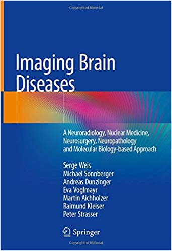
Imaging Brain Diseases: A Neuroradiology, Nuclear Medicine, Neurosurgery, Neuropathology and Molecular Biology-based Approach 1st ed. 2019 Edition
by Serge Weis (Author), Michael Sonnberger (Author), Andreas Dunzinger (Author), Eva Voglmayr (Author), Martin Aichholzer (Author), Raimund Kleiser (Author), Peter Strasser (Author)
Hardcover: 2284 pages
Publisher: Springer; 1st ed. 2019 edition (January 21, 2020)
Language: English
ISBN-10: 3709115434
ISBN-13: 978-3709115435
For Download Please Contact Us :
Price : 25$
ادرس اینترنتی کتاب از اشپرینگر :
https://www.amazon.com/Imaging-Brain-Diseases-Radiological-Neuropathological/dp/3709115434
دانلود رایگان کتاب Imaging Brain Diseases A Neuroradiology Nuclear Medicine Neurosurgery Neuropathology Molecular Biology-based Approach
دانلود کتاب Decommissioning Forecasting and Operating Cost Estimation
This book illustrates in a unique way the most common diseases affecting the human nervous system using different imaging modalities derived from radiology, nuclear medicine, and neuropathology. The features of the diseases are visualized on computerized tomography (CT)-scans, magnetic resonance imaging (MRI)-scans, nuclear medicine scans, surgical intraoperative as well as gross-anatomy and histology preparations. For each disease entity, the structural changes are illustrated in a correlative comparative way based on the various imaging techniques. The brain diseases are presented in a systematic way allowing the reader to easily find the topics in which she or he is particularly interested. In Part 1 of the book, the imaging techniques are described in a practical, straightforward way. The morphological built-up of the normal human brain and its vascular supply are presented in Part 2. The chapters of the subsequent Parts 3 to 10 deal with the following diseases involving the nervous system including: hemodynamic, vascular, infectious, neurodegenerative, demyelination, epilepsy, trauma and intoxication, and tumors.
The authors incite the clinician to see the cell, the tissue, the organ, the disorder by enabling him to recognize brain lesions or interpreting histologic findings and to correlate this knowledge with molecular biologic concepts. Thus, this book bridges the gap between neuro-clinicians, neuro-imagers and neuro-pathologists. The information provided will facilitate the understanding of the disease processes in the daily routine work of neurologists, neuroradiologists, neurosurgeons, neuropathologists, and all allied clinical disciplines.
دانلود کتاب تصویربرداری از مغز مغز A Neuroradiology هسته ای پزشکی هسته ای Neuropathology Neuropathology رویکرد مبتنی بر زیست شناسی مولکولی
این کتاب به روشی منحصر به فرد شایع ترین بیماری های مؤثر بر سیستم عصبی انسان را با استفاده از روش های مختلف تصویربرداری به دست آمده از رادیولوژی ، پزشکی هسته ای و نوروپاتولوژی نشان می دهد. ویژگی های این بیماری ها بر روی توموگرافی کامپیوتری (CT) – وسایل نقلیه ، تصویربرداری با رزونانس مغناطیسی (MRI) ، اسکن پزشکی پزشکی هسته ای ، جراحی حین جراحی و همچنین آماده سازی ناخالص آناتومی و بافت شناسی مشاهده می شود. برای هر موجودیت بیماری ، تغییرات ساختاری به روش مقایسه ای مبتنی بر تکنیک های مختلف تصویربرداری نشان داده شده است. بیماری های مغز به صورت منظم ارائه می شود و به خواننده امکان می دهد موضوعاتی را که در آن به ویژه علاقه مند است ، به راحتی پیدا کند. در قسمت 1 کتاب ، تکنیک های تصویربرداری به روشی عملی و ساده بیان شده است. ساختار مورفولوژیکی مغز طبیعی انسان و تأمین عروق آن در بخش 2 ارائه شده است. فصل های قسمتهای بعدی 3 تا 10 با بیماری های زیر درگیر سیستم عصبی از جمله: همودینامیک ، عروقی ، عفونی ، عصبی ، دفع زدا ، و صرع ، تروما و مسمومیت و تومورها.
نویسندگان با استفاده از این پزشک ، پزشک را به دیدن سلول ، بافت ، اندام و اختلال در ایجاد ضایعات مغزی یا تفسیر یافته های بافت شناسی و همبستگی این دانش با مفاهیم بیولوژیک مولکولی تحریک می کنند. بنابراین ، این کتاب شکاف بین پزشکان متخصص مغز و اعصاب ، تصویرگرهای اعصاب و آسیب شناسان را ایجاد می کند. اطلاعات ارائه شده ، درک فرآیندهای بیماری را در کار روزمره روان شناسان ، اعصاب و روان ، جراحان مغز و اعصاب ، مغز و اعصاب و کلیه رشته های بالینی متفقین تسهیل خواهد کرد.