دانلود کتاب Pediatric Dentistry Infancy through Adolescence 7th edition
خرید ایبوک Pediatric Dentistry نسخه 7ام سال 2026
برای دانلود ایبوک Pediatric Dentistry: Infancy through Adolescence, 7th edition و خرید کتاب دندانپزشکی کودکان (نوزادی تا نوجوانی) نواک ویرایش هفتم سال 2026 بر روی کلید خرید در انتهای صفحه کلیک کنید. پس از اتصال به درگاه پرداخت هزینه و تکمیل مراحل خرید، لینک دانلود کتاب ایمیل می شود.این ایبوک در فرمت PDF اورجینال و به زبان انگلیسی ارسال می شود.نسخه الکترونیکی کتاب قابلیت کپی برداری copy و Paste دارد.
در صورتی که نیاز به پی دی اف هر کتابی از انتشارات Saunders دارید با گیگاپیپر مکاتبه کنید.
خرید کتاب Pediatric Dentistry: Infancy through Adolescence 7 th E-Book – PDF
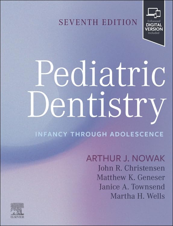
Pediatric Dentistry: Infancy through Adolescence, 7th edition Original pdf
by Arthur J. Nowak DMD MA (Editor), John R. Christensen John R. Christensen DDS MS (Ped Dent) MS (Ortho) (Editor), Matthew Geneser DDS (Editor), Janice Alisa Townsend DDS MS (Editor), Martha H. Wells DMD MS (Editor)
Publisher : Saunders
Publication date : September 1, 2026
Edition : 7th
Language : English
Print length : 656 pages
ISBN-10 : 0443246173
ISBN-13 : 978-0443246173
Send email to download :
Price : 35$
https://www.vitalsource.com/en-uk/products/pediatric-dentistry-arthur-j-nowak-v9780443250392

https://www.amazon.com/Pediatric-Dentistry-Infancy-through-Adolescence/dp/0443246173
دانلود رایگان کتاب پدیاتریک دنتیستری Pediatric Dentistry در فرمت پی دی اف اورجینال و اصلی
Provide superior oral care to children of all ages! Pediatric Dentistry: Infancy through Adolescence, 7th Edition, provides comprehensive coverage of oral care for infants, children, teenagers, and children with disabilities and chronic health conditions. The text covers examination, diagnosis, and treatment planning, as well as topics such as the prevention of oral disease, orthodontics, pulp therapy, oral trauma, and behavioral management. This edition includes new treatments, techniques, and evidence-based guidelines, plus expanded case studies in the enhanced ebook included with every print purchase. From a team of accomplished authors and contributors led by Arthur J. Nowak, this book also prepares you for success on the INBDE and other exams.
- Comprehensive coverage of current trends and challenges emphasizes the prevention of oral diseases and reflects pediatric dentistry as it is practiced today
- Evidence-based focus includes topics such as local and systemic diseases, pediatric physiology, cariology, pain control, and medical emergencies ― all core concepts for both the dental student and the practicing dentist treating children
- More than 1,000 full-color photos and illustrations show oral conditions and treatments
- Diverse contributor pool includes expert perspectives from all over the world
- Enhanced ebook version, included with every new print purchase, features videos and case studies with review questions, plus digital access to all text, figures, and references, with the ability to search, customize content, make notes and highlights, and have content read aloud on a variety of devices
- NEW content covers emerging concepts within pediatric dentistry, including management of unique patient populations, updates in technologies and materials, and pediatric dentistry for the practicing general dentist
- NEW! Updates on caries risk assessment in children reflect the evolution of evidence-based oral health care
- NEW and expanded case studies are included in the enhanced ebook
دانلود ایبوک کتاب دندانپزشکی اطفال: نوزادی تا نوجوانی، ویرایش هفتم (زبان اصلی)
Pediatric Dentistry
Infancy through Adolescence
7th Edition
Author(s)Arthur J. Nowak
Publisher : Published by Saunders Copyright© 2027
Print ISBN: 9780443246173
مراقبتهای دهانی برتر را برای کودکان در تمام سنین فراهم کنید! دندانپزشکی کودکان: از نوزادی تا نوجوانی، ویرایش هفتم، پوشش جامعی از مراقبتهای دهانی برای نوزادان، کودکان، نوجوانان و کودکان دارای معلولیت و بیماریهای مزمن ارائه میدهد. متن شامل معاینه، تشخیص و برنامهریزی درمان و همچنین مباحثی مانند پیشگیری از بیماریهای دهان، ارتودنسی، درمان پالپ، ترومای دهان و مدیریت رفتاری است. این ویرایش شامل درمانها، تکنیکها و دستورالعملهای جدید مبتنی بر شواهد، به علاوه مطالعات موردی گسترده در کتاب الکترونیکی پیشرفتهای است که با هر خرید نسخه چاپی ارائه میشود. این کتاب که توسط تیمی از نویسندگان و مشارکتکنندگان برجسته به رهبری آرتور جی. نواک نوشته شده است، شما را برای موفقیت در آزمون INBDE و سایر آزمونها نیز آماده میکند.
پوشش جامع روندها و چالشهای فعلی، بر پیشگیری از بیماریهای دهان تأکید دارد و دندانپزشکی کودکان را آنطور که امروزه انجام میشود، منعکس میکند. تمرکز مبتنی بر شواهد شامل موضوعاتی مانند بیماریهای موضعی و سیستمیک، فیزیولوژی کودکان، پوسیدگیشناسی، کنترل درد و فوریتهای پزشکی است – همه مفاهیم اصلی برای دانشجوی دندانپزشکی و دندانپزشک شاغل در درمان کودکان. بیش از ۱۰۰۰ عکس و تصویر تمام رنگی، شرایط و درمانهای دهان را نشان میدهند. مجموعه متنوعی از مشارکتکنندگان شامل دیدگاههای متخصصان از سراسر جهان است. نسخه الکترونیکی پیشرفته، که با هر خرید نسخه چاپی جدید ارائه میشود، شامل ویدیوها و مطالعات موردی با سوالات مروری، به علاوه دسترسی دیجیتال به تمام متن، شکلها و منابع، با قابلیت جستجو، سفارشیسازی محتوا، یادداشتبرداری و برجستهسازی و امکان خواندن محتوا با صدای بلند در دستگاههای مختلف است.
محتوای جدید، مفاهیم نوظهور در دندانپزشکی کودکان، از جمله مدیریت جمعیتهای منحصر به فرد بیمار، بهروزرسانیها در فناوریها و مواد و دندانپزشکی کودکان را برای دندانپزشک عمومی شاغل پوشش میدهد.
جدید! بهروزرسانیها در مورد ارزیابی خطر پوسیدگی در کودکان، منعکسکننده تکامل مراقبتهای بهداشتی دهان مبتنی بر شواهد است.
مطالعات موردی جدید و گسترده در کتاب الکترونیکی پیشرفته گنجانده شده است.
کتاب دنداپزشکی کودکان نواک ویرایش 7 هفتم
فهرست مطالب ایبوک کتاب Pediatric Dentistry اثر Arthur J. Nowak
- Pediatric Dentistry: INFANCY THROUGH ADOLESCENCE
- Cover image
- Title page
- Table of Contents
- Copyright
- Contributors
- Foreword
- Preface
- Video contents
- List of Illustrations
- List of Tables
- PART 1. Dynamics of Change
- 1. Importance of pediatric dentistry
- Prevention and diagnosis
- Behavior guidance
- Treatment options
- Dental disease emphasis and systemic disease
- Contemporary practice and care of children
- This edition
- References
- 2. Craniofacial growth—conception through adolescence
- Physical, craniofacial, and dental changes: From birth through adolescence
- Growth and development: Birth to 3 years
- Growth and development: 3 to 6 years
- Growth and development: 6 to 12 years
- Growth and development: Adolescence
- References
- 3. Cognitive, emotional, and social development—infancy to age 3
- Cognitive changes
- Emotional and social changes
- Developmental surveillance
- Screen time for infants and toddlers
- References
- 4. Cognitive, emotional, and social development—ages 3 to 6 years
- Screen time
- Parenting styles
- References
- 5. Cognitive, emotional, and social development—ages 6 to 12 years
- Cognitive changes
- Emotional changes
- Social changes
- Gender identity
- Peers, friendships, and bullying
- Smartphones, internet, and social media
- Effects of parenting
- COVID-19 pandemic effects
- Problematic media exposure
- Summary
- References
- 6. Cognitive, emotional and social development—adolescence
- Cognitive changes
- Emotional changes
- Social changes
- Bullying
- Mental health and emotional well-being
- Gender identity
- Nutrition and physical fitness
- Risky behaviors
- Tobacco and nicotine products
- Sexual activity
- Social media and internet use
- Teen dating violence and human trafficking
- Drug and alcohol use
- Summary
- Acknowledgments
- References
- 7. Principles of anatomy and physiology for the pediatric dentist
- Introduction
- Head and neck
- Nervous system
- Brain
- Cranial nerves
- Respiratory system
- Cardiovascular system
- Conclusion
- References
- PART 2. A Clinical Guide to Treating the Pediatric Patient
- 8. Infant oral health and the toddler: Ages 0 to 3
- Introduction
- Infant oral health: Definition, rationale, and goals
- Break the cycle of early childhood caries
- Disrupt the acquisition of harmful microflora
- Manage the risk/benefit of habits
- Establish a dental home
- Impart optimal fluoride protection
- Arming the parents through anticipatory guidance
- Normal findings
- Preparing for your patient
- Office readiness for infant oral health
- Elements of the infant oral health visit
- Special considerations
- Growth and development treatment planning
- Nonnutritive sucking habits
- References
- 9. The preschool child: Ages 3 to 6
- Normal findings
- Preparing for your patient
- Information gathering
- Behavior guidance
- Clinical examination
- Radiographic examination
- Caries risk assessment
- Prevention
- Toothpaste recommendations
- Nonfluoride preventive adjuncts
- Nutrition advice
- Restorative considerations
- Minimally invasive dentistry
- Referrals
- Special considerations
- Oral habits
- Occlusion
- Space maintenance
- References
- 10. The school-aged child: Ages 6 to 12
- Normal findings
- Preparing for your patient
- Information gathering
- Behavior guidance
- Clinical examination
- Radiographic examination
- Prevention
- Restorative considerations
- Referrals
- Special considerations
- Management of hypomineralized molars
- Pulp therapy for primary teeth
- Pulp therapy for young permanent teeth
- Management and prevention of traumatic dental injuries
- Orthodontic management in mixed dentition
- References
- 11. Adolescent oral health
- Introduction
- Normal findings
- Preparing for your patient
- Patient history
- Behavior and coping skills
- Clinical examination
- Radiographic examination
- Prevention
- Caries risk assessment
- Periodontal risk assessment
- Assessing for third molars
- Restorative considerations
- Transitioning to the adult dental home
- Special considerations
- Dental bleaching
- Orthodontic management of permanent dentition
- Temporomandibular joint disorder
- Sports dentistry
- Eating disorders
- Human papillomavirus vaccinations
- Treating the pregnant adolescent patient
- Intraoral and perioral piercing
- Intraoral tattoos
- Substance misuse
- References
- PART 3. Fundamentals
- 12. Cariology
- Introduction
- Brief overview of caries
- Importance of understanding cariology in pediatric dentistry
- Historical introduction
- Timeline of dentistry with pivot points
- Historical findings by Miller, Black, Keyes, and Stephan
- Caries microbiota and oral biofilm
- Composition and role of oral microbiota in caries development
- Key bacterial species involved
- Research on the ecologic shifts and dysbiosis leading to caries
- Role of saliva
- Saliva’s protective functions against caries
- Components of saliva and their roles
- Impact of saliva flow and composition on caries risk
- Remineralization and regenerative technology
- Remineralization
- Transmission of caries
- Mechanisms of caries transmission (vertical and horizontal)
- Role of caregivers and siblings in transmission
- Preventive measures to reduce transmission risk
- Risk assessments
- Updated caries risk assessment models
- Factors influencing caries risk (e.g., genetic, environmental, behavioral)
- Integration of risk assessments into clinical practice
- Emerging diagnostic methodologies
- Early caries lesion detection
- Salivary diagnostics
- Biofilm/microbiota biomarkers
- Evidence-based dentistry and ethical use of diagnostic technology: Role of artificial intelligence
- Antimicrobials and vaccines
- Use of antimicrobial agents in caries prevention and management
- Current and emerging vaccines targeting cariogenic bacteria
- Future directions in cariology research
- Implications for pediatric dental practice
- References
- 13. Prevention in pediatric dentistry in today’s healthcare
- Introduction
- The whole patient
- The family, community, culture, and society: Social determinants of health
- The system: Its benefits and limitations
- Medicaid
- Targeted prevention in private pediatric dentistry
- Summary
- References
- 14. Fluoride and nonfluoride caries preventive agents
- Introduction
- Fluoride
- Fluoride mechanism of action
- Metabolism
- Fluoride modalities
- Side effects of fluoride exposure
- Nonfluoride preventive agents
- Chlorhexidine
- Povidone iodine
- Arginine
- Xylitol
- Casein phosphopeptide-amorphous calcium phosphate
- Hydroxyapatite
- References
- Case study: Fluoride toxicity
- 15. Pit and fissure sealants: Scientific and clinical rationale
- Epidemiology of pit and fissure caries
- Sealant effectiveness
- Current sealant utilization
- Types of sealants
- Properties of sealant materials
- Color of resin sealants
- Filler content of resin sealants
- Fluoride-releasing resin sealants
- Hydrophilic resin sealant
- Glass ionomer
- Hybrid
- The necessity of sealants
- Caries risk assessment
- Sealant and fluoride varnish
- Age at placement
- Which teeth should be sealed?
- Diagnosing occlusal caries
- Sealing over incipient caries
- Preparing the tooth for a sealant
- Factors affecting sealant success
- Hypomineralized enamel
- Auxiliary application
- Four-handed delivery
- School-based sealant programs
- Other uses for sealant
- Interproximal dental sealant
- Sealing restorations
- Preventing white spot lesions
- Sealant safety
- Future advances
- Summary
- References
- Case study: Rubber dam split dam technique and resin dental sealant placement
- Case study: Glass ionomer sealant placement
- 16. Pediatric diet and nutrition for oral health
- Normal nutrition
- Growth and development
- Oral health and disease
- Dental caries
- Periodontal disease
- Tooth wear
- Outcome of oral disease
- Dietary counseling
- Conclusion
- References
- 17. Radiology for the pediatric dental patient
- Introduction and general concepts
- Intraoral imaging modalities
- Periapical radiograph
- Bitewing radiograph
- Extraoral imaging modalities
- Panoramic radiograph
- Cephalometric radiograph
- Cone beam computed tomography (CBCT)
- Considerations across pediatric age groups
- The transitional years: 6 to 12 years
- Adolescence
- References
- 18. Pediatric pharmacology
- Antimicrobial classification and use
- Microbiology
- Chemotherapeutic spectrum of activity
- Bacteriostatic versus bactericidal agents
- Categories of antibiotic use
- Antimicrobial mechanisms of action
- Inhibition of cell wall synthesis
- Inhibition of protein synthesis
- Suppression of DNA synthesis
- Alteration of cell membrane permeability
- Adverse effects of antimicrobials
- Allergic reactions
- Photosensitivity reactions
- Long QT interval syndrome
- Drug-drug interactions
- Antibacterial agents
- Penicillins
- Cephalosporins (cephams)
- Clindamycin
- Macrolides
- Metronidazole
- Fluoroquinolones
- Antifungal agents
- Polyenes
- Azole antifungals
- Antiherpetic agents
- Acyclovir
- Valacyclovir
- Special considerations in the pediatric patient
- Barriers to compliance
- Accurate dosing
- Unpalatable medications
- Clinical use of antimicrobials in pediatric dentistry
- Bacterial infections
- Viral infections
- Fungal infections
- Antimicrobial prophylaxis
- References
- 19. Differential diagnosis of oral lesions and developmental anomalies
- Suggested reading
- Case study: Orofacial gr anulomatosis
- 20. Anomalies of the developing dentition
- Developmental controls and environmental interactions
- Anomalies of tooth number
- Hyperdontia
- Hypodontia
- Anomalies of size
- Microdontia
- Macrodontia
- Anomalies of tooth morphology
- Crown size and morphology
- Root size and morphology
- Dental tissue composition and structural defects
- Environmental defects altering tooth color
- Hereditary enamel defects
- Hereditary dentin defects
- Hereditary cementum defects
- Anomalies of tooth eruption
- Primary failure of eruption
- Summary
- References
- Case study: Oral pathology—anomalies of teeth
- References
- 21. Periodontal problems in children and adolescents
- Gingivitis
- Localized juvenile spongiotic gingival hyperplasia
- Gingival enlargement
- Chronic inflammatory gingival enlargement
- Plaque-induced, drug-influenced gingival enlargement
- Anatomical problems
- Development and defects of the attached gingiva
- Frena
- Periodontitis
- Grade C periodontitis
- Necrotizing ulcerative gingivitis/periodontitis
- Systemic diseases and conditions with associated periodontal problems
- Diabetes
- Down syndrome
- Leukocyte adhesion deficiency
- Neutropenia
- Papillon-Lefèvre syndrome
- Histiocytosis
- Leukemia
- Periodontal examination of children
- References
- Case study: Laser frenectomy
- References
- 22. Space maintenance in the primary and mixed dentition
- General considerations
- Loss of primary incisors
- Loss of primary canines
- Loss of primary molars
- Additional considerations
- Space-maintaining appliances
- Fixed unilateral appliances
- Fixed bilateral appliances
- Removable appliances
- Fabrication and laboratory considerations
- Summary
- References
- 23. Oral habits
- Nonnutritive sucking habits
- Behavior modification techniques
- Reminder therapy
- Appliance therapy
- Treatment interventions
- Atypical swallowing
- Mouth breathing
- Bruxism
- Lip habits
- Nail biting
- Summary
- References
- Case study: Diagnosis, treatment planning, and correction of a thumb habit
- Diagnosis and treatment planning
- Treatment
- 24. Orthodontic treatment in the primary dentition
- Facial examination
- Overall facial pattern
- Positions of the maxilla and mandible
- Vertical facial relationships
- Lip position
- Facial symmetry
- Intraoral examination
- Occlusal evaluation
- Alignment
- Skeletal problems
- Transverse growth modification
- Anteroposterior growth modification
- Vertical growth modification
- Dental problems
- Potential alignment and space problems
- Missing permanent teeth
- Tooth size discrepancies
- Alignment problems
- Anteroposterior dental problems
- Transverse dental problems
- Vertical dental problems
- Crowding problems
- References
- Case study: Treating ectopic eruption of first permanent molars
- Case study: Orthodontics for the child with disabilities
- Behavior
- Patient’s ability to tolerate the appliance
- Patient’s ability to cooperate with the treatment plan
- Stability of results
- References
- 25. Orthodontic treatment in the permanent dentition
- Determination of developmental status
- Head and neck examination
- Facial examination
- Intraoral examination
- Periodontal evaluation
- Related hard and soft tissue problems
- Occlusal evaluation
- Treatment planning and treatment for orthodontic problems
- Skeletal problems
- Dental problems
- References
- 26. Behavior guidance of the pediatric dental patient
- Understanding the child patient: Review of child development
- Cognitive development
- Learning theory
- Temperament
- Coping
- Factors influencing child behavior
- Dental fear
- Demographics
- Environment
- Pain
- Influence of the parent
- Setting the stage for successful behavior guidance
- The dental office
- Preparing the child for the dental visit
- Scheduling
- The dentist and the dental team
- Patient assessment
- Parents in the operatory
- Setting parent expectations
- Review of behavior guidance techniques
- Basic behavior guidance
- Additional techniques to consider
- Behavior guidance for the child with previous negative dental experiences
- Advanced behavior guidance techniques
- Mouth props in dentistry for children
- Documenting behavior and use of behavior management techniques
- Role of behavior management in society
- Changes in society and parenting
- Changing parental perspectives of behavior management
- Third-party reimbursement
- Informed consent
- Conclusion
- References
- Case study: Prepared parents, p repared children, positive coping
- Case description
- Key considerations
- Approach
- Case study: Protective stabilization
- 27. Behavior guidance-pharmacologic
- Introduction
- Levels of sedation
- Minimal and moderate sedation
- Deep sedation
- General anesthesia
- Minimal and moderate versus deep sedation: A critical question
- Rescue
- The guidelines as the standard of care for minimal and moderate sedation
- Routes of administration
- Inhalational route
- Practical use of inhalation sedation techniques
- Oral route
- Intranasal route
- Intramuscular route
- Intravenous route
- Intravenous access
- Preoperative assessment
- Common pharmacologic agents used for sedation
- Antianxiety agents
- Analgesic agents
- Balanced technique
- Monitors
- Pulse oximeter
- Precordial stethoscope
- Capnography
- Recovery and discharge
- The future
- Summary
- References
- Case study: Considerations for sedating the pediatric patient
- 28. Assessment and management of pain in the pediatric patient
- The impact and management of pain
- Definition of pain
- Pain neuroscience
- Pain processing
- Disrupting pain signaling
- Theories of pain
- Gate control
- Conditioned pain modulation
- Neuromatrix
- Biopsychosocial
- Peripheral and central nervous system effects on pain perception and control
- Cognitive elements of pain perception
- Emotional elements of pain perception
- Pediatric pain assessment
- Pain control with analgesics
- Nonopioid analgesics
- Acetaminophen
- Nonsteroidal antiinflammatory drugs
- Aspirin
- Opioid analgesics
- Dosing schedule
- Multimodal therapy
- Pain control with local anesthesia
- Mechanisms of action
- Local anesthetic agents
- Excretion of local anesthetics
- Local anesthetic properties
- Potency
- Onset time
- Duration
- Regional technique
- Onset
- Duration
- Other factors
- Central nervous system reactions
- Cardiovascular system reactions
- Operator technique
- Topical anesthesia
- Needle selection
- Injection technique
- Complications of local anesthesia
- Alternative anesthesia systems
- Buffered local anesthesia
- Intranasal anesthetic spray
- Computer-controlled intraligamentary anesthesia
- Computer-assisted intraosseous anesthesia
- Vibrotactile devices
- Cryo-vibrotactile devices
- Conclusion: Local anesthetics
- References
- Case study: Local anesthesia
- Failure of mandibular block
- Use of a computerized device for anesthetizing maxillary incisors
- References
- 29. Dental materials
- Bases and liners
- Calcium hydroxide
- Zinc oxide–eugenol
- Glass ionomer cement
- Dentin-bonding agents
- Restorative materials
- Amalgam
- Resin-based composite
- Glass ionomer
- Compomers
- Luting cements
- Stainless steel crowns
- Monolithic zirconia
- References
- 30. Restorative dentistry for the primary dentition
- Anatomical considerations of primary teeth
- Instrumentation and caries removal
- Isolation in pediatric restorative dentistry
- Rubber dam
- Alternative isolation systems
- Restoration of primary molars
- Amalgam use in primary molars
- Adhesive materials in primary molars
- Adhesive systems for primary teeth
- General principles for intracoronal restoration of primary posterior teeth
- Restoration of primary incisors and canines
- Class III restorations
- Class V restorations
- Full coronal coverage of incisors
- Prosthetic replacement of primary anterior teeth
- References
- Case study: Pediatric zirconia crowns
- 31. Restorative dentistry for the adolescent
- Fundamentals of material selection
- Fundamentals of clinical technique
- Restorations for fractured anterior teeth
- Clinical technique: Tooth fragment
- Clinical technique: Class IV restoration
- Restoration of diastemas
- Clinical technique
- Restoration of discolored teeth
- Vital bleaching
- Minimally invasive treatments
- Veneers
- Strip crowns
- Restorations for posterior teeth
- Fundamentals of material selection
- Resin infiltration
- Composite resin
- Amalgam
- Resin-modified glass ionomer
- Stainless steel crowns
- Enamel hypomineralization
- Restoring the hypomineralized molar
- Managing rampant caries
- References
- Case study: Management of the hypomineralized molar
- Case study: Resin infiltration
- Description of resin infiltration
- Case study: Management of the uncomplicated crown fracture
- 32. Pulp therapy for the primary dentition
- Histology
- The pulp-dentin complex
- Dentinogenesis in healthy state
- Dentinogenic response to injury
- Dentin-pulp complex response to stimuli in primary teeth
- Clinical pulpal diagnosis
- Medical history
- Extra- and intraoral examination
- Pain characteristics
- Sensibility tests
- Preoperative diagnosis of deep caries lesions
- Operative diagnosis
- Trauma
- Correlation between histopathologic status of the pulp and deep caries
- Pulp treatment procedures
- Vital pulp therapy for normal pulp/reversible pulpitis
- Pulp therapy for irreversible pulpitis or necrotic pulp
- Summary
- References
- Case study: Primary tooth mineral trioxide aggregate (MTA) pulpotomy
- 33. Pulp therapy for the young permanent dentition
- The dentin-pulp complex concept
- Reactions to caries and operative procedures
- Clinical pulpal diagnosis
- Patient history
- Clinical examination
- Pain history
- Radiographic examination
- Direct pulp evaluation
- Vital pulp therapy for permanent teeth
- Terminology for caries removal
- Indirect pulp treatment
- Protective liner
- Direct pulp capping
- The pulpotomy procedure
- Nonvital pulp treatment for immature teeth
- Apexification
- Long-term apexification with calcium hydroxide
- One-visit apexification with mineral trioxide aggregate
- Regenerative endodontics
- Nonvital pulp treatment for young mature teeth
- Root canal treatment in young mature permanent teeth: Special considerations
- References
- Case study: Permanent molar pulpotomy
- References
- Case study: Management of complicated crown fractures in permanent teeth
- References
- 34. Pediatric oral surgery: Principles and techniques
- Introduction to pediatric oral surgery: Principles and practices
- Scope of pediatric oral surgery
- Preoperative assessment
- Medical and dental history
- Risk assessment
- Physical examination
- Radiographic assessment
- Behavioral and psychological considerations
- Informed consent and patient/parent education
- Extractions in pediatric patients
- Indications for extraction
- Armamentarium for pediatric extractions
- Techniques for extraction
- Postoperative care
- Management of bleeding
- Third molar extractions: Current philosophies, indications, and timing
- Current treatment philosophies
- Indications for extraction
- Timing of extraction
- Risks of extraction
- Odontogenic infections in pediatric patients
- Types and causes of odontogenic infections
- Diagnosis and clinical signs
- Treatment approaches
- Trauma management in pediatric patients
- Surgical interventions
- Management of facial fractures
- Long-term follow-up and monitoring
- Pharmacomanagement in pediatric oral surgery
- Pain management
- Intraoperative pain management
- Postoperative pain management
- Prescription analgesics
- Sedation and general anesthesia
- Antibiotics and other medications
- Special procedures in pediatric oral surgery
- Biopsy and pathology
- Suturing in pediatric oral surgery
- Management of impacted teeth
- Implants in pediatric patients
- Autotransplantation
- Surgical management of ankylosed teeth
- Complications in pediatric oral surgery
- Prevention and risk mitigation
- Postoperative care and follow-up in pediatric oral surgery
- Future directions in pediatric oral surgery
- 3D imaging
- Laser surgery
- Research and clinical trials
- Conclusion
- References
- Case study: Extraction of primary molar
- Reference
- PART 4. Special Considerations
- 35. Medical emergencies
- Medical emergencies in the pediatric dental office
- Morbidity
- Mortality
- “Swiss cheese model” of accident causation
- Prevention of medical emergencies
- History and physical examination
- Medical consultation
- Patient monitoring
- Preparing for office emergencies
- Personal preparation
- Staff preparation
- Backup medical assistance
- Office preparation: Emergency equipment
- Management of medical emergencies in the dental office
- Position
- Circulation (“C”)
- Airway (“A”)
- Breathing (“B”)
- Definitive therapy (“D”)
- Common medical emergencies
- Allergic reactions
- Asthma (acute bronchospasm)
- Cardiac symptoms (acute coronary syndrome)
- Cardiac arrest
- Diabetic emergencies
- Hyperventilation syndrome
- Seizures
- Syncope
- Sedation-related emergencies
- Candidates for sedation in pediatric dentistry
- Rescue skills for deeper levels of sedation
- Airway management and perioperative respiratory adverse events
- Maintaining competency
- Knowledge of sedative agents
- Recommendations
- References
- 36. Children with disabilities
- Overview of disability
- Disability
- Developmental disability
- Intellectual disability
- Physical disability
- Sensory disability
- Chronic health conditions
- Medically complex
- Children with disabilities
- Biopsychosocial model of disability
- Neurodiversity
- Children with disabilities in the dental setting
- Assessment
- Common developmental disabilities
- Risk of social isolation
- Parents
- Siblings
- Implications for dentists
- Ableism
- Ableism in language
- Person-first and identity-first language
- Barriers to care
- Systemic barriers to care
- Clinical barriers to care
- Barriers to evidence-based dentistry
- Oral health of children with disabilities
- Oral hygiene
- Caries risk
- Oral hygiene and caries prevention strategies
- Trauma risk and management
- Behavior guidance
- Best practices
- Strengths-based approach
- Universal design
- Patient- and family-centered care
- Sensory-adapted dental environment
- Communication
- Social stories
- Desensitization
- Transition
- Conclusions
- References
- Case study: Children with suspected autism spectrum disorder
- References
- 37. Congenital genetic disorders and syndromes
- Basic genetic concepts
- Molecular basis of disease
- Inheritance patterns
- Autosomal dominant
- Autosomal recessive
- X-linked
- Chromosomal anomalies
- Multifactorial inheritance
- Nontraditional inheritance
- Dentist as dysmorphologist
- Ocular anomalies
- Auricular anomalies
- Anomalies of the mouth and oral region
- Dental anomalies
- Syndromes/disorders with craniofacial anomalies
- Down syndrome
- Ectodermal dysplasia
- Cleidocranial dysplasia
- Williams-Beuren syndrome
- Fragile X syndrome
- Osteogenesis imperfecta
- Treacher collins syndrome
- Van der woude syndrome
- Isolated (nonsyndromic) orofacial clefts
- Hypophosphatasia
- Apert syndrome
- Crouzon syndrome
- Genetic testing
- Ethical, legal, and social implications of the human genome project
- References
- Case study: Assessing inherited disorders
- Case
- 38. Cleft lip and palate
- Cleft lip and palate team
- Anatomy
- Classification of cleft lip and palate
- Causes of cleft lip and palate
- Challenges and anomalies associated with cleft lip and palate
- Infant orthopedics
- An overview of the timeline of interventions
- Role of the dentist
- References
- Case study: Presurgical nasoalveolar molding
- Case 1
- Case 2
- 39. Dental and oral considerations for inherited and acquired diseases
- Inherited coagulation disorders
- Hemophilia
- Von willebrand disease
- Dental considerations for patients with inherited coagulation disorders
- Anemia
- Dental considerations for patients with anemia
- Sickle cell disorders
- Dental considerations for patients with sickle cell disorders
- Glucose-6-phosphate dehydrogenase deficiency
- Dental considerations for patients with G6PD deficiency
- Gastroenterology
- Gastroesophageal reflux disease
- Dental considerations for patients with gastroesophageal reflux disease
- Celiac disease
- Dental considerations for patients with celiac disease
- Inflammatory bowel disease
- Crohn disease
- Ulcerative colitis
- Dental considerations for patients with inflammatory bowel disease
- Diabetes mellitus
- Dental considerations for patients with diabetes mellitus
- Epilepsy
- Dental considerations in patients with epilepsy
- Asthma
- Dental considerations in patients with asthma
- Cystic fibrosis
- Dental considerations in patients with cystic fibrosis
- Congenital heart disease
- Pediatric infective endocarditis
- Pediatric cancer
- Hematopoietic stem cell transplantation
- Immunotherapy and targeted therapy
- Oral complications from cancer therapy
- Dental considerations before, during, and after cancer treatment
- Treatment planning considerations
- During active cancer treatment
- After cancer treatment
- Oral hygiene, diet, and caries prevention throughout cancer treatment
- References
- Case study: Cyanotic congenital heart disease
- 40. Introduction to dental trauma: Managing traumatic injuries in the primary dentition
- Etiology and epidemiology of trauma in the primary dentition
- Classification of injuries to teeth
- History
- Medical history
- History of the dental injury
- Oral health quality of life
- Clinical examination
- Extraoral examination
- Intraoral examination
- Radiographic examination
- Emergency care
- Injuries to the hard dental tissues
- Injuries to the supporting tissues
- Information and instructions for parents
- Pathologic sequelae of trauma to the teeth
- Reversible pulpitis
- Infection of the periodontal ligament
- Coronal discoloration
- Rapidly progressing root resorption
- Replacement external root resorption
- Pulp canal obliteration
- Complications after intrusion
- Complications after avulsion
- Injuries to developing permanent teeth
- Soft tissue injuries
- Summary
- References
- 41. Managing traumatic injuries in the young permanent dentition
- Etiology and epidemiology of trauma in the young permanent dentition
- Classification of injuries to young permanent teeth
- History
- Clinical examination
- Pathologic sequelae of traumatized teeth
- Treatment of traumatic injuries to the permanent dentition
- Enamel fractures
- Enamel and dentin fractures
- Fractures involving the pulp
- Posterior crown fractures
- Root fractures
- Managing sequelae to dental trauma
- Treating luxation injuries in the permanent dentition
- References
- Summary
- References
- Case study: Splinting for an avulsed tooth
- Preparation for splint placement
- References
- 42. Sports dentistry
- Introduction
- Definition of sports dentistry
- Risk of orofacial injuries in sports
- Dental trauma in sports
- Prevention of dental injuries in sports
- Face shields
- Mouthguards
- Types of injuries
- Dental injuries
- Soft tissue injuries
- Temporomandibular joint injuries
- Bone injuries
- Concussions
- Mouthguard types
- History of mouthguards
- Future directions for sports dentistry
- Sports nutrition
- Oral health as a part of total health and implications for athletic performance
- Intraoral appliances for enhanced athletic performance
- Summary
- References
- Case study: Mouthguards and management of sports-related dental trauma
- 43. Abuse, neglect, bullying, and human trafficking
- Introduction
- Ethical and legal obligation
- Decision making
- Physical abuse
- Bite marks on the skin
- Sexual abuse
- Medical child abuse
- Bullying
- Human trafficking
- Dental neglect
- Abuse and neglect in children with special healthcare needs
- Teen dating violence (intimate partner violence)
- Adverse childhood exposures
- Documentation and protocols
- Conclusion
- References
- 44. Ensuring accessible care for all—dental public health
- Introduction
- Definition of dental public health
- Role of the individual practitioner
- Access to care
- Dental participation in medicaid
- The pediatric dental benefit within the affordable care act
- Federally qualified health centers
- Prenatal and early childhood oral health: The role of nondental professionals and public health programs
- Project head start
- Special medicaid dental programs
- School-based dental care
- References
- Case study: Addressing sexual identity in pediatric dental patients
- 45. Temporomandibular disorders
- Temporomandibular disorders in children and adolescents
- Temporomandibular joint anatomy and function
- Temporomandibular joint growth and development
- Introduction to temporomandibular disorders
- Etiology of temporomandibular disorders
- Diagnosis and treatment of temporomandibular disorders
- Screening assessment for temporomandibular disorders
- Comprehensive assessment for temporomandibular disorders
- Psychosocial assessment
- Imaging and temporomandibular disorders
- Management of temporomandibular disorders
- Idiopathic condylar resorption
- References
- Case study: Chronic jaw pain
- Background
- Interview
- Clinical examination
- Radiographic examination
- Problem list
- Diagnosis
- Treatment plan and recommendations
- 46. Management of missing teeth in a growing patient
- Introduction and background
- Diagnosis and assessment
- Clinical examination and records
- Treatment planning
- Multidisciplinary collaboration
- Treatment options
- Removable appliances
- Fixed appliances
- Orthodontic space closure
- Tooth autotransplantation
- Patient selection criteria
- Donor tooth selection
- Recipient site preparation
- Dental implants
- Extraction of permanent first molars
- Summary
- References
- 47. Sleep-disordered breathing
- Etiology of pediatric obstructive sleep apnea
- Anatomical factors
- Craniofacial development
- Genetic factors
- Environmental factors
- Clinical presentation of sleep-disordered breathing in children
- Screening for sleep-disordered breathing
- Medical and family history review
- Cognitive, behavioral, and developmental assessment
- Screening tools and questionnaires
- Clinical oral and craniofacial examination
- Interdisciplinary collaboration and referrals
- Diagnostic tool for pediatric obstructive sleep apnea
- Polysomnography
- Home sleep apnea testing
- Treatment plans for pediatric sleep-disordered breathing1
- Adenotonsillectomy
- Medical management
- Positive airway pressure therapy
- Orthodontic and dentofacial interventions
- Myofunctional therapy
- Growth modification techniques and proposed guidelines for sleep-disordered breathing in children
- Preschool stage (infancy to 5 years: Primary dentition)
- Early childhood (ages 6 to 9: Early mixed dentition)
- Middle childhood (ages 10 to 11: Late mixed dentition)
- Adolescence (ages 12 to 15: Permanent dentition)
- Postadolescence (ages 16 to 18: Permanent dentition)
- Conclusion
- References
- 48. Tethered oral tissues
- Definitions and terminology
- Epidemiology
- Classification of frenal attachments
- Maxillary labial frenum
- Lingual frenum
- Conception to age three
- Breastfeeding
- Oral hygiene and caries risk
- Surgical intervention for prevention
- The primary dentition years: Three to six years
- Speech development
- Airway and breathing considerations
- The transitional years: Six to twelve years
- Maxillary labial frenum and midline diastema
- Frenula and orthodontic treatment
- Social/mechanical issues
- Surgical intervention
- Definitions
- Surgical technique
- Postsurgical care
- Complications
- Special considerations: Craniofacial anomalies, syndromic and nonsyndromic conditions
- Conditions associated with abnormal frenal attachments
- Tethered oral tissues in patients with cleft lip and palate
- References
- Index